


Mucicarmine Stain
Mucicarmine Stain Service: Detect Epithelial Mucin with Clarity and Contrast
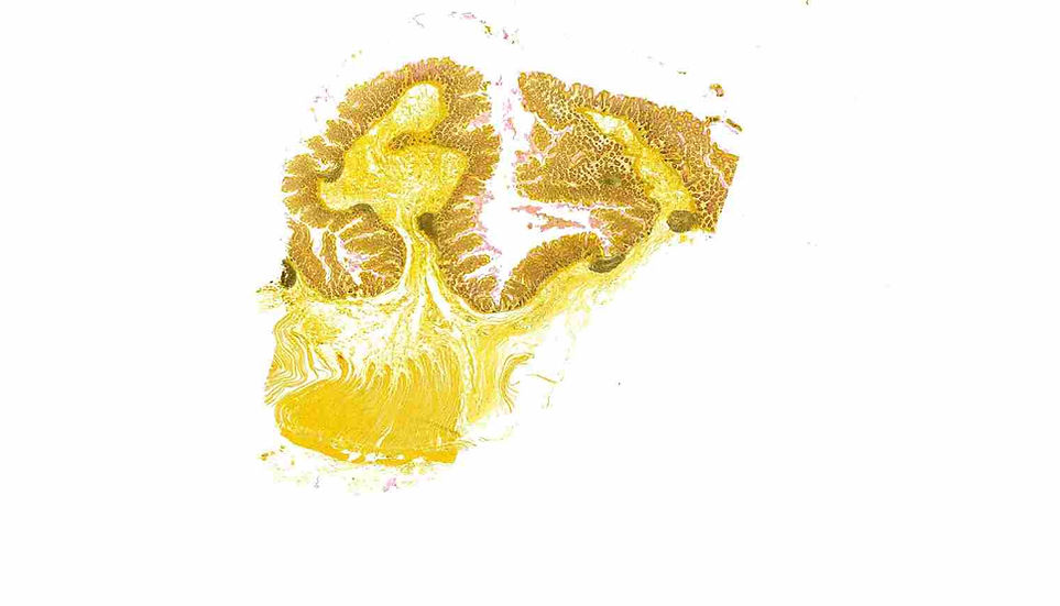
At iHisto, we offer precise Mucicarmine staining to detect epithelial mucins in tissue sections. This stain is essential for identifying mucin-producing tumors, detecting Cryptococcus neoformans, and evaluating glandular epithelium in gastrointestinal and pulmonary samples.
🧪 Overview
The Mucicarmine stain is a targeted histochemical technique that highlights acidic mucins in epithelial tissues. It provides strong contrast for identifying:
Adenocarcinomas of the GI tract, lung, or salivary glands
Cryptococcal infections through capsule staining
Mucin dynamics in inflammation or metaplasia
iHisto’s refined protocol ensures consistent mucin detection with excellent specificity.
⚙️ How Mucicarmine Staining Works
Mucicarmine binds to acidic epithelial mucins via an aluminum-carmine complex, producing a rose to deep red coloration. A nuclear counterstain such as hematoxylin or metanil yellow provides contrast.
Color Results:
Mucin → pink to deep red
Nuclei → blue to black
Background / cytoplasm → yellow (if metanil yellow is used)
This allows for confident identification of mucin-producing cells and structures.
🧬 Step-by-Step Staining Process
Fixation & Sectioning
FFPE tissue is cut at 4–6 µm.Deparaffinization & Hydration
Slides are cleared and rehydrated to prepare for staining.Mucicarmine Staining
The primary mucin stain is applied to target acidic mucins.Counterstaining
Hematoxylin and optional metanil yellow are applied for nuclei and background contrast.Dehydration & Coverslipping
Slides are cleared, dehydrated, and sealed for analysis or archiving.
🔬 Applications of Mucicarmine Staining
The Mucicarmine stain is commonly used in:
Adenocarcinoma diagnostics – GI, pulmonary, salivary origin
Fungal identification – highlights Cryptococcus capsule
Differentiating mucinous tumors – primary vs metastatic origin
Barrett’s esophagus / intestinal metaplasia
IBD pathology – mucin depletion or overproduction
This stain is frequently used alongside H&E, PAS, and Alcian Blue for comprehensive mucin characterization.
✅ Why Choose iHisto
✅ Sharp, selective staining of epithelial mucins
✅ Validated for human and animal tissues
✅ Compatible with special stain panels
✅ Whole-slide scanning and digital delivery included
✅ Optional pathologist review and annotation
We support research in oncology, infectious disease, and gastrointestinal pathology with reliable mucin-focused histology.
❓ FAQs
What does Mucicarmine stain detect?
It detects epithelial mucins and highlights the polysaccharide capsule of Cryptococcus neoformans.Is it used in tumor classification?
Yes. It confirms mucinous adenocarcinomas and helps differentiate primary from metastatic tumors.Can it be used in infectious disease research?
Absolutely. It is a reliable tool for identifying Cryptococcus in tissue sections.
Do you provide digital slides?
Yes — we offer high-resolution whole-slide scanning with cloud or encrypted offline delivery.
📩 Request a Quote or Consultation
Whether you're diagnosing mucinous tumors, confirming fungal infections, or analyzing epithelial changes, iHisto delivers high-quality Mucicarmine staining tailored for your research.
👉 Request a Quote or email info@ihisto.io