


Verhoeff Van Gieson Stain
Verhoeff Van Gieson (VVG) Stain: Clear Differentiation of Elastic Fibers and Collagen
At iHisto, we provide Verhoeff Van Gieson (VVG) staining to distinguish elastic fibers, collagen, and muscle/cytoplasmic elements in tissue — essential for evaluating fibrosis, vascular remodeling, and connective tissue integrity.
🧪 Overview
VVG staining combines Verhoeff’s elastic stain with Van Gieson’s collagen counterstain, offering multi-component visualization in a single slide. It is particularly valuable in:
Vascular pathology – Aorta, arteries, aneurysms
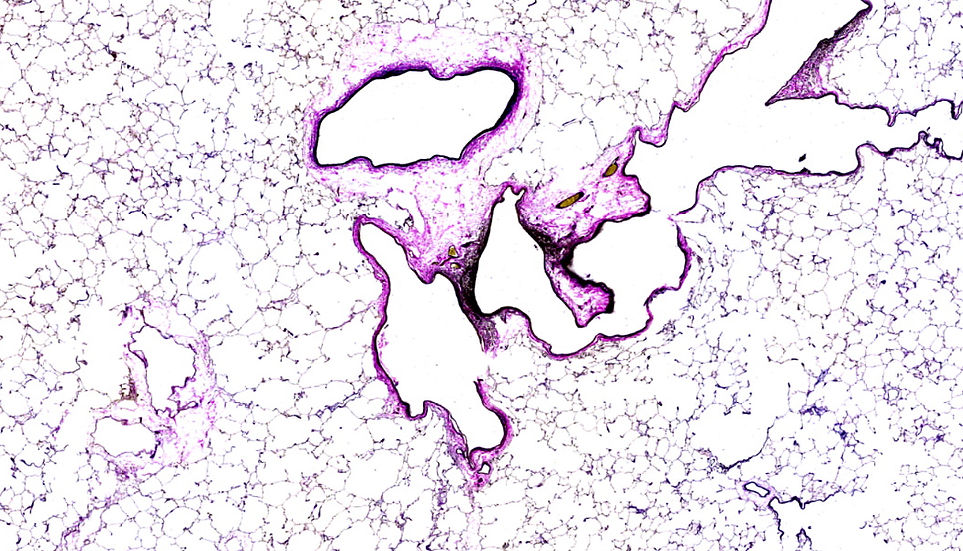
Pulmonary research – Fibrosis, alveolar elastin
Dermatopathology – Elastosis, aging, or connective tissue diseases
Liver & tumor studies – Fibrosis and stromal architecture
iHisto’s optimized protocol delivers consistent, high-contrast results for both preclinical models and human specimens.
⚙️ How VVG Staining Works
VVG involves two key reactions:
Verhoeff’s Stain: Hematoxylin, ferric chloride, and iodine bind to elastic fibers, turning them deep black
Van Gieson Counterstain: Acid fuchsin stains collagen red, and picric acid stains muscle/cytoplasm yellow
Color Results:
Elastic fibers → black
Collagen → red
Muscle/cytoplasm → yellow
Nuclei → dark blue to black
This allows precise tissue architecture evaluation, especially in disease models affecting elastin and ECM.
🧬 Step-by-Step Staining Process
Fixation & Sectioning – FFPE tissue cut at 4–6 µm
Deparaffinization & Hydration – Slides rehydrated into water
Verhoeff’s Elastic Staining – Elastic fibers stained black
Differentiation – Ferric chloride enhances contrast
Van Gieson Counterstaining – Collagen turns red; muscle/cytoplasm yellow
Dehydration & Mounting – Slides sealed for microscopy and archiving
🔬 Applications of Verhoeff Van Gieson Staining
VVG is widely used in histology to assess fibrotic remodeling and elastic fiber structure.
Common use cases:
Aortic pathology – Elastic laminae damage, medial degeneration
Pulmonary fibrosis – Loss of alveolar elastin
Skin biopsies – Solar elastosis, aging changes
Liver fibrosis models – Complements Trichrome/Reticulin staining
Tumor analysis – Evaluate stromal ECM and desmoplasia
Whether you're studying ECM remodeling or tissue degeneration, VVG offers unparalleled clarity of elastic and collagen components.
✅ Why Choose iHisto for VVG Staining
✅ High-contrast, reproducible staining of elastin and collagen
✅ Compatible with human and rodent tissue
✅ Suitable for vascular, dermal, pulmonary, and liver studies
✅ Optional pathologist review and interpretation
✅ Digital slide scanning with HistoCloud delivery
We support clinical research, CRO studies, and academic institutions with customized VVG staining solutions.
❓ FAQs
What does Verhoeff Van Gieson stain detect?
Elastic fibers (black), collagen (red), muscle/cytoplasm (yellow), and nuclei (blue to black).Is this stain used for aortic or pulmonary pathology?
Yes — VVG is commonly used to assess aortic elastic laminae and pulmonary alveolar structure.Can it be applied to both rodent and human samples?
Absolutely. Our validated workflow is suitable for clinical specimens and preclinical research tissues.Do you provide slide digitization?
Yes — we offer whole-slide scanning, accessible via secure cloud or encrypted storage.
📩 Request a Quote or Consultation
Need high-contrast visualization of elastic fibers and collagen? At iHisto, our Verhoeff Van Gieson staining service supports vascular, pulmonary, dermal, and fibrotic pathology — with rapid turnaround, expert processing, and digital accessibility.
👉 Request a Quote or email info@ihisto.io